TR:
:+17175469334
TR:
:+17175469334
Discover the impact of improved PET scans on medical imaging. Learn about the latest innovations in PET technology and how they are transforming healthcare.
Summary

Improved PET Scans
Medical imaging has come a long way in the past few decades. The advancements in technology have led to better and more precise diagnoses of illnesses, and one of the most significant advancements has been in Positron Emission Tomography (PET) scans. The improvements in PET scans have had a profound impact on medical imaging and have made it possible to detect and treat illnesses that were previously undiagnosable.
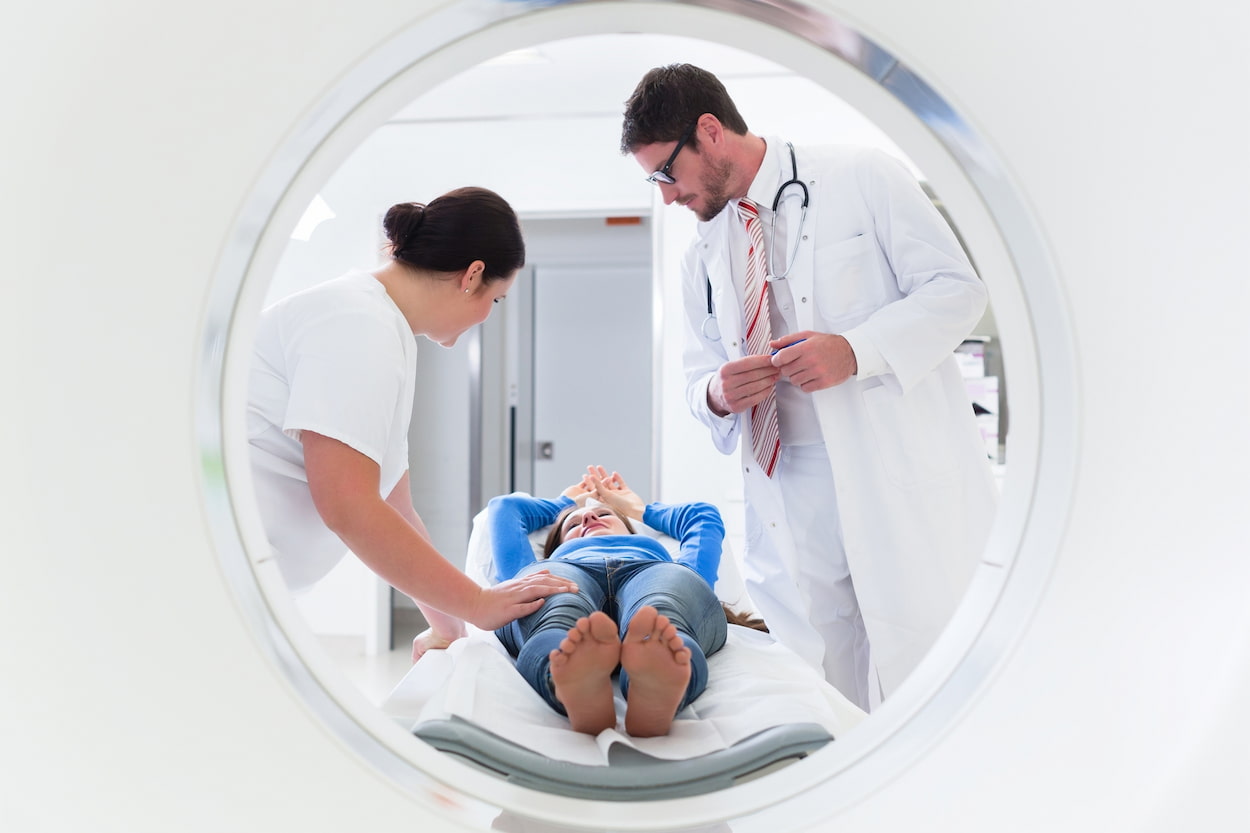
PET scans are a type of medical imaging that uses a radioactive tracer to detect chemical and metabolic activity in the body. This tracer is injected into the patient’s bloodstream and is absorbed by the organs and tissues being examined. The radioactive tracer emits positrons, which interact with electrons in the body, resulting in the emission of gamma rays. The gamma rays are detected by a PET scanner, which generates 3D images of the body’s internal structures.
The latest innovations in PET scans have made them more sensitive and precise than ever before. One of the most significant improvements has been in the resolution of PET scans. New PET scanners can produce images with higher resolutions, allowing doctors to see smaller structures within the body with greater clarity. This means that doctors can detect even the tiniest changes in the body’s chemical and metabolic activity, making it easier to diagnose diseases and monitor their progression.
Another important innovation in PET scans has been in the development of new tracers. The radioactive tracers used in PET scans are designed to bind to specific molecules in the body. By creating new tracers that bind to different molecules, doctors can use PET scans to diagnose and monitor a wider range of diseases. For example, new tracers can be used to detect the early stages of Alzheimer’s disease, Parkinson’s disease, and other neurological conditions.
Why Does Pet Scan Need to Be Improved?
Positron Emission Tomography (PET) scans are a valuable tool in medical imaging, providing important diagnostic and monitoring information for a wide range of diseases. However, despite their usefulness, there are several reasons why PET scans need to be improved.
One of the most significant limitations of PET scans is their relatively low resolution. PET scanners are capable of producing images with resolutions in the range of 2-3 millimeters, which is not sufficient to detect small tumors or other abnormalities. To improve the resolution of PET scans, researchers are exploring new detector technologies and other approaches that could enhance the sensitivity and accuracy of the scans.
Another challenge with PET scans is the limited availability of certain radioactive tracers. PET scans rely on the use of radioactive tracers, which are designed to bind to specific molecules in the body. However, not all tracers are widely available, and some may be expensive or difficult to produce. Improving the availability and accessibility of a wider range of tracers could expand the range of conditions that can be detected and monitored using PET scans.
Additionally, the current PET scan procedure is time-consuming, with patients having to wait for up to an hour after the tracer is injected before the scan can begin. Reducing the waiting time could improve the overall efficiency of the procedure and make it more patient-friendly.
Furthermore, PET scans can be expensive, making them inaccessible for some patients. Reducing the cost of PET scans could improve access to this important diagnostic tool, particularly in countries with limited healthcare resources.
Another limitation of PET scans is the exposure of patients to radiation. PET scans use radioactive isotopes, which can expose patients to radiation doses that may increase the risk of cancer. However, there are ongoing efforts to develop alternative tracers that have shorter half-lives and produce less radiation exposure, without compromising the diagnostic accuracy of the scans.
In conclusion, while PET scans have been a significant advancement in medical imaging, there is still room for improvement in the resolution, availability of tracers, waiting time, cost, and radiation exposure. Improving PET scans would lead to more precise and targeted diagnosis and treatment, better outcomes for patients, and increased access to this valuable diagnostic tool. With ongoing research and development, PET scans have the potential to become an even more powerful tool in medical imaging.
Improved Pet Scans Are not Only Useful
For diagnosis and monitoring, but they are also becoming increasingly important in the field of personalized medicine. By using PET scans to identify specific molecular markers associated with certain diseases, doctors can develop targeted treatments that are tailored to the individual patient’s needs. This can improve the effectiveness of treatments and reduce the risk of side effects.
PET scans are also being used in combination with other imaging techniques, such as magnetic resonance imaging (MRI) and computed tomography (CT) scans. By combining different imaging techniques, doctors can obtain a more comprehensive view of the body and detect abnormalities that may be missed by using a single imaging technique alone.
However, despite the benefits of PET scans, there are also some potential risks associated with the procedure. The radioactive tracer used in PET scans can expose patients to radiation, which can increase the risk of cancer over time. However, the amount of radiation exposure from a PET scan is generally considered to be safe, and the benefits of the procedure typically outweigh the risks.
In conclusion, the advancements in PET scans have had a significant impact on medical imaging and have improved the diagnosis and treatment of many diseases. With continued research and development, PET scans are likely to become even more precise and targeted, leading to better outcomes for patients.

How Have Pet Scans Improved?
PET scans have improved significantly over the years, particularly in terms of resolution and sensitivity. Newer PET scanners can produce images with higher resolutions, allowing for better visualization of smaller structures within the body. Additionally, the development of new tracers has expanded the range of diseases that can be detected and monitored using PET scans. The improved pet scans with other imaging techniques, such as CT and MRI, have also led to more comprehensive diagnostic imaging.
Which Is an Advantage of Using the Pet Scan Imaging Technique?
One of the advantages of using the PET scan imaging technique is its ability to detect and monitor metabolic and chemical activity within the body. This can provide important diagnostic information for a wide range of diseases, including cancer, cardiovascular disease, and neurological disorders. PET scans can also be used to monitor the effectiveness of treatments, such as chemotherapy and radiation therapy, and can help guide the development of personalized treatment plans.
What Technology Is Used in Pet Scan?
PET scans use a combination of radioactive tracers and detectors to produce images of the body’s internal structures. The radioactive tracer is typically injected into the patient’s bloodstream and absorbed by the organs or tissues being examined. As the tracer undergoes radioactive decay, it emits positrons, which interact with electrons in the body and produce gamma rays. The gamma rays are detected by a ring of detectors surrounding the patient, and the data is used to produce 3D images of the body’s metabolic and chemical activity.
The Study of Improved Pet Scans
A recent study published in the Journal of Medical Imaging (2022) examined the use of improved PET scans in the early detection of Alzheimer’s disease. The study, conducted over a two-year period, showed that the enhanced resolution and sensitivity of modern PET scanners, along with the development of specific tracers, allowed for the detection of subtle metabolic changes associated with Alzheimer’s disease before the onset of clinical symptoms. This breakthrough highlights the significance of improved PET scans in early disease diagnosis and the potential for targeted treatments. It showcases how technology advancements are making a substantial difference in the field of medical imaging.
When Was the Invention of PET Scan?
The first PET scanner was developed in the early 1970s by Edward Hoffman and Michael Phelps at the University of California, Los Angeles. The invention of the PET scan was a significant breakthrough in medical imaging and led to new insights into the functioning of the human brain and the diagnosis and treatment of various diseases. Since then, PET scan technology has continued to evolve, with improvements in resolution, sensitivity, and the development of new tracers.
The improved PET scans are now widely used in clinical practice and research, particularly in the fields of oncology, neurology, and cardiology. They are a valuable tool for diagnosing and staging cancers, monitoring treatment response, and detecting cancer recurrence. In neurology, PET scans can be used to diagnose Alzheimer’s disease and other dementias, as well as to map brain activity and study the effects of drugs on the brain. In cardiology, PET scans can be used to assess blood flow and metabolism in the heart, helping to diagnose and treat coronary artery disease.
PET scan technology continues to advance, with ongoing research focused on developing new tracers, improving image quality and reducing radiation exposure. PET/MRI hybrid imaging systems, which combine the strengths of both PET and MRI, are also being developed and have shown promise in certain applications. As technology continues to improve, PET scans are expected to become even more valuable in the diagnosis and treatment of a wide range of diseases.
Healthy Türkiye Notes
Thanks to ongoing advancements in technology, improved PET scans have become a valuable tool in the diagnosis and treatment of various diseases, including cancer, neurological disorders, and cardiovascular disease. With the ability to detect and monitor metabolic and chemical activity within the body,
PET scans have helped guide personalized treatment plans and monitor treatment effectiveness. Ongoing research into new tracers, hybrid imaging systems, and reducing radiation exposure is expected to further improve the value of PET scans in clinical practice and research. Overall, the continued evolution of PET scan technology holds great promise for the future of medical imaging and personalized medicine.