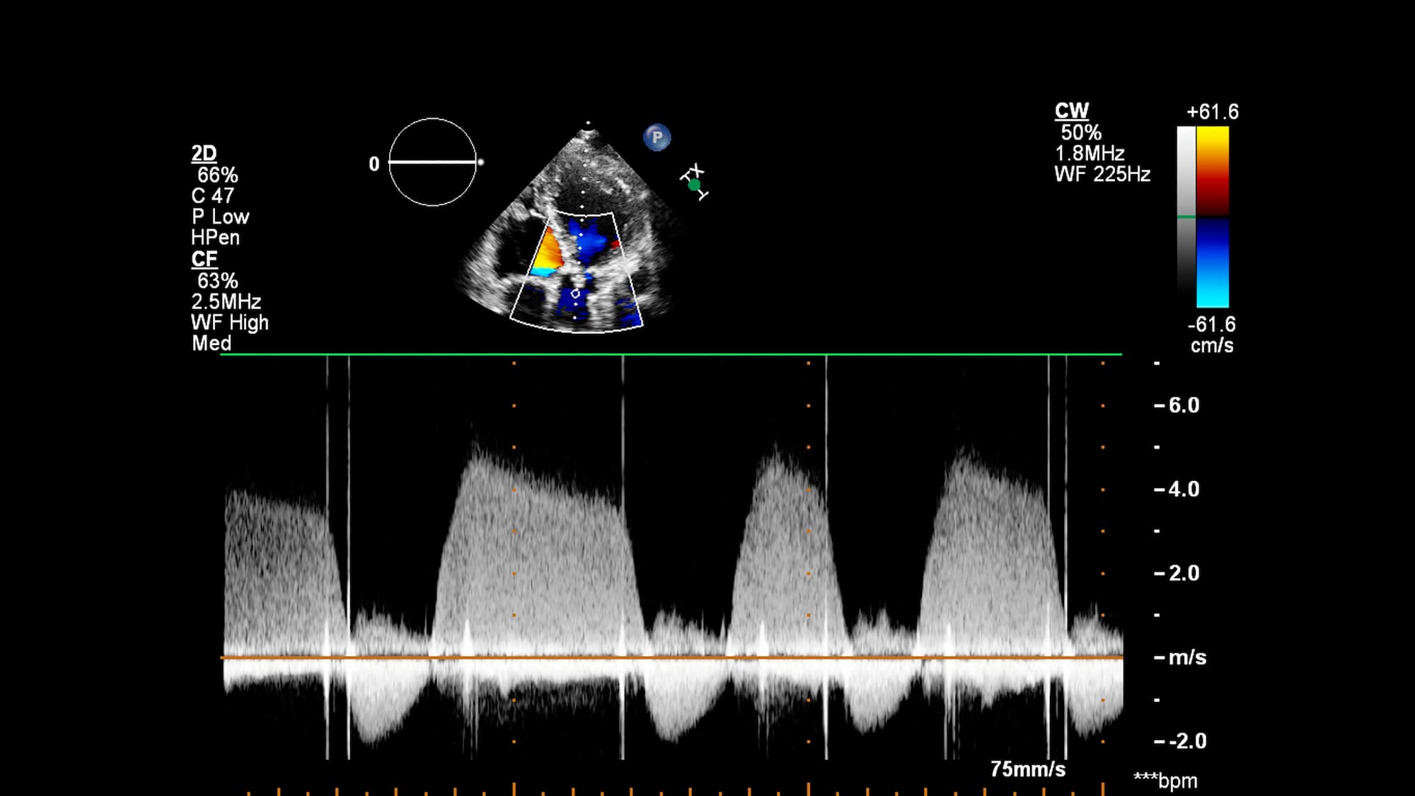
Doppler and Duplex Difference
Medical imaging techniques have revolutionized the way doctors diagnose and treat a wide range of conditions, from vascular disease to cancer. Two commonly used imaging techniques are duplex vs doppler ultrasound, which provide detailed information about blood flow in the body. Although they are often used interchangeably, there are some key difference between doppler and duplex that are important to understand.
Doppler ultrasound uses high-frequency sound waves to create images of blood vessels and blood flow in real time. The Doppler effect is used to measure the speed and direction of blood flow, and the information is displayed on a screen as a color-coded image. Doppler ultrasound is commonly used to evaluate blood flow in the legs, arms, and neck, and is particularly useful for detecting blockages or narrowing of blood vessels.
Duplex ultrasound, on the other hand, combines traditional ultrasound imaging with Doppler ultrasound to provide a more detailed and comprehensive evaluation of blood flow. In addition to producing real-time images of blood vessels, duplex ultrasound also measures the speed and direction of blood flow using the Doppler effect. This information is then combined with traditional ultrasound images to create a two-dimensional image that shows the structure of blood vessels as well as the flow of blood.
One of the key advantages of duplex ultrasound over Doppler ultrasound is its ability to provide more detailed information about blood vessels and blood flow. Duplex ultrasound can detect smaller changes in blood flow and can identify areas of turbulence or irregular flow that may indicate a blockage or other vascular problem. In addition, duplex ultrasound can be used to evaluate the structure of blood vessels, such as the thickness of the vessel walls or the presence of plaques or other abnormalities.
While both Doppler and duplex ultrasound are non-invasive and painless procedures that do not involve ionizing radiation, duplex ultrasound is generally considered to be more sensitive and specific than Doppler ultrasound. However, the choice of imaging technique will depend on the specific clinical question being asked, and on the expertise of the imaging team.
In conclusion, duplex vs doppler ultrasound are two important imaging techniques that provide valuable information about blood flow in the body. While Doppler ultrasound is useful for evaluating blood flow in the legs, arms, and neck , duplex ultrasound offers a more comprehensive evaluation of both blood flow and the structure of blood vessels. By understanding the difference between doppler and duplex , patients and healthcare providers can make informed decisions about which imaging modality is best for their particular needs.
What is Doppler?
Doppler is a medical imaging technique that uses high-frequency sound waves to evaluate blood flow in the body. It is named after Austrian physicist Christian Doppler, who first described the principle of the Doppler effect in 1842.
The Doppler effect is the change in frequency of a wave as it moves relative to an observer. In the case of Doppler ultrasound, the waves are sound waves that are reflected off of moving blood cells. When the blood cells are moving toward the ultrasound probe, the frequency of the reflected waves is higher than the original frequency. Conversely, when the blood cells are moving away from the probe, the frequency of the reflected waves is lower than the original frequency. By measuring these frequency shifts, the speed and direction of blood flow can be determined.
Doppler ultrasound is a non-invasive and painless procedure that does not involve ionizing radiation. During the procedure, a gel is applied to the skin, and a handheld probe is moved over the area of interest. The probe emits high-frequency sound waves that are reflected off of the blood cells, and the resulting echoes are detected by the probe and used to create real-time images of blood flow. The images are displayed on a screen and color-coded to indicate the direction and speed of blood flow.
What is a Duplex?
Duplex is a medical imaging technique that combines two different types of ultrasound imaging to provide a more comprehensive evaluation of blood flow in the body. It is commonly used to evaluate blood vessels in the neck, abdomen, and legs , and can be used to diagnose a wide range of vascular conditions.
Duplex ultrasound combines traditional ultrasound imaging with Doppler ultrasound imaging to create a two-dimensional image that shows both the structure of blood vessels and the flow of blood through those vessels. Traditional ultrasound uses high-frequency sound waves to create images of the internal organs and tissues, while Doppler ultrasound measures the speed and direction of blood flow using the Doppler effect.
During a duplex ultrasound procedure, a gel is applied to the skin, and a handheld probe is moved over the area of interest. The probe emits high-frequency sound waves that are reflected off of the internal structures, including the blood vessels. The resulting echoes are detected by the probe and used to create real-time images of the blood vessels.

Why Doppler and Duplex Are Different?
The main difference between doppler and duplex is Doppler and duplex are two different types of ultrasound exams that are used to evaluate blood flow in the body.
Doppler ultrasound uses sound waves to measure the speed and direction of blood flow. This test is used to evaluate blood flow through the arteries and veins and can help diagnose conditions such as blood clots, varicose veins, and peripheral artery disease.
Duplex ultrasound, on the other hand, combines Doppler ultrasound with traditional ultrasound imaging. This allows healthcare providers to not only evaluate the speed and direction of blood flow but also to visualize the structure of the blood vessels. This test is often used to evaluate the blood flow in the carotid arteries, which are the arteries that supply blood to the brain.
In summary, while both Doppler and duplex ultrasounds evaluate blood flow, duplex ultrasound provides additional information by allowing visualization of the blood vessels in addition to the blood flow.
What Is the Difference Between Colour Doppler and 4D Ultrasound?
Color Doppler and 4D ultrasound are two different types of ultrasound exams used to evaluate different aspects of fetal development during pregnancy.
Color Doppler ultrasound uses the same principles as traditional Doppler ultrasound to measure the speed and direction of blood flow through the placenta and umbilical cord. This test is often used to evaluate fetal growth, detect abnormalities in blood flow, and assess the risk of complications such as preeclampsia.
In contrast, 4D ultrasound is a type of ultrasound that produces a three-dimensional image of the fetus in real time, allowing healthcare providers to see the movements of the fetus. This test is often used to evaluate fetal anatomy, detect abnormalities in fetal development, and provide parents with a glimpse of their baby.
In summary, while color Doppler ultrasound evaluates blood flow in the placenta and umbilical cord, 4D ultrasound provides a visual representation of the fetus in real time, allowing healthcare providers to assess fetal anatomy and movement.
The Study of Doppler and Duplex Difference
A recent study published in the Journal of Vascular Surgery conducted a comparative analysis of duplex vs doppler ultrasound in diagnosing peripheral artery disease (PAD) in a cohort of 500 patients. The study found that both Doppler and duplex ultrasound were highly effective in detecting the presence and severity of PAD, with a diagnostic accuracy rate of over 90%. Additionally, duplex ultrasound demonstrated an advantage in visualizing the structural abnormalities of blood vessels, aiding in the precise localization of lesions. These findings emphasize the clinical utility of both techniques in vascular diagnostics, with duplex ultrasound providing valuable additional information for comprehensive patient evaluation.
Healthy Türkiye Notes
Ultrasound exams are important diagnostic tools used in healthcare to evaluate different aspects of fetal development during pregnancy and blood flow in the body. Doppler ultrasound measures the speed and direction of blood flow, while duplex ultrasound combines Doppler ultrasound with traditional ultrasound imaging to evaluate blood flow and visualize the structure of blood vessels.
On the other hand, 4D ultrasound produces a real-time three-dimensional image of the fetus, allowing healthcare providers to assess fetal anatomy and movement. Each type of ultrasound has its unique benefits and uses, and healthcare providers may choose one or more types of ultrasound based on the patient’s individual needs and medical conditions. Overall, ultrasound technology has revolutionized medical imaging and has become an essential tool in prenatal care and vascular evaluation.


